Introduction to Leukemia & Acute Leukemia
Presentation44 slidesB.Pharm, Pharm.D
Loading…
About this presentation
Notes on Leukemia, types of Leukemia, epidemiology, pathophysiology of Leukemia.
- leukemia
- acute leukemia
- hematological malignancy
- epidemiology
- pathophysiology
Text of these slides
- Slide 1. LEUKEMIA Prepared by Dr. Ayesha Fatima Pharm D Assistant Professor Dept of Pharmacy Practice ACUTE LEUKEMIA
- Slide 2. Leukemia is a heterogeneous group of hematologic malignancies characterized by the uncontrolled proliferation of abnormal blood-forming cells (blasts) in the bone marrow. These immature cells: • Accumulate in the bone marrow • Inhibit normal blood cell production • Leukemic blasts may also infiltrate extramedullary tissues such as: 1. Lymph nodes 2. Liver and spleen 3. Skin 4. Kidneys 5. Central nervous system (CNS) 6. Testes leading to anemia , neutropenia , and thrombocytopenia . } 🠀
- Slide 3. Type Cell Line Clinical Course Predominant Age Group Acute Lymphoblastic Leukemia (ALL) Lymphoid Acute Children Acute Myeloid Leukemia (AML) Myeloid Acute Adults Chronic Lymphocytic Leukemia (CLL) Lymphoid Chronic Older Adults Chronic Myeloid Leukemia (CML) Myeloid Chronic Adults Leukemia is classified by two main criteria: 1. By Cell Lineage (Origin): • Lymphoid (Lymphocytic) → derived from lymphoid progenitors • Myeloid (Myelogenous) → derived from myeloid progenitors 2. By Clinical Course: • Acute: Rapid onset and aggressive course, immature blasts dominate • Chronic: Slow progression, cells are more mature and differentiated The Four Major Types of Leukemia:
- Slide 7. • Acute leukemia is fatal within weeks to months if untreated • Leukemia accounts for ~1.5% of all cancers diagnosed annually • Causes about 2% of all cancer-related deaths • >90% of leukemia cases occur in adults • In adults with acute leukemia: 80% AML, 20% ALL • In children: 75% ALL, 25% AML India Ranks 3rd in Blood Cancer Age Group Leukemia Type Notes < 20 years ALL (High), AML (less) Leukemia is the most common cancer in this age group (27% of all childhood cancers) 20–39 years Leukemia Leading cause of cancer-related death > 40 years AML, CLL, CML Less common but more likely to occur in adults and elderly EPIDEMIOLOGY
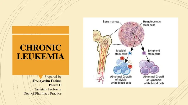
- Slide 8. 1. Origin: All blood cells arise from pluripotent hematopoietic stem cells (HSCs) in the bone marrow. 2. Differentiation Pathways: a. Myeloid stem cells → Erythrocytes, platelets, neutrophils, eosinophils, basophils, monocytes. b. Lymphoid stem cells → T lymphocytes, B lymphocytes, natural killer (NK) cells. 3. Processes Involved: Hematopoiesis involves proliferation, differentiation, and maturation of cells under tight genetic control. Normal Hematopoiesis
- Slide 10. 1. Clonal Origin of Leukemia • Leukemia begins with a single abnormal (leukemic) cell. • This cell undergoes genetic mutations → gains the ability to proliferate uncontrollably. • Results in a monoclonal population of abnormal blasts (myeloblasts or lymphoblasts). 2. Imbalance in Cell Development a. There is a failure to maintain balance between: a) Proliferation (cell multiplication) b) Differentiation (maturation into functional blood cells) b. Cells are arrested at an immature stage → accumulate as blasts. c. Normal hematopoiesis is suppressed, leading to: i. Anemia (↓ RBCs) ii. Thrombocytopenia (↓ platelets) iii. Neutropenia (↓ mature WBCs) PATHOPHYSIOLOGY OF LEUKEMIA
- Slide 11. 3. Site and Stage of Origin Varies by Type a. Acute Myeloid Leukemia (AML): • Usually originates from pluripotent stem cell or early myeloid precursor. b. Acute Lymphoblastic Leukemia (ALL): • Arises from lymphopoietic stem cell or early lymphoid precursor. • Leads to accumulation of lymphoblasts. 4. Crowding Out Effect • Proliferating blasts overcrowd bone marrow, reducing space for normal cells. • Leads to: • Decreased production of functional RBCs, WBCs, and platelets. • Clinical signs: fatigue, infections, bleeding tendencies. 5. Not Due to Rapid Proliferation Alone • Leukemic cells may not proliferate faster, but: • They survive longer (↓ apoptosis). • They may secrete factors that: • Inhibit normal cell proliferation/differentiation. • Protect themselves from apoptosis.
- Slide 12. 6. Genetic and Molecular Abnormalities • Leukemogenesis involves multiple genetic alterations, including: a. Proto-oncogene activation → forms oncogenes that drive proliferation. b. Differentiation arrest → loss of signals needed for proper maturation. c. Loss of tumor suppressor genes → unchecked cell division. d. Impaired apoptosis → cells avoid programmed cell death, resist therapy. 7. Affected Cellular Processes • Signal transduction • RNA transcription • Cell cycle regulation • Differentiation and maturation • Apoptosis (programmed cell death) Flowchart: Normal HSC (genetic mutation) leukemic clone loss of differentiation + ↑ survival blast accumulation bone marrow failure + systemic manifestations
- Slide 13. ACUTE LEUKEMIA
- Slide 14. Acute leukemia results from genetic mutations in hematopoietic stem cells, leading to: 1. Unregulated proliferation of immature white blood cells (blasts) 2. Blocked differentiation into mature blood cells 3. These mutations may be: a. Acquired (somatic) → due to environmental exposures, drugs, infections, etc. b. Inherited (germline) → due to genetic syndromes Etiology and Risk Factors of Acute Leukemia
- Slide 15. Clinical Presentation Manifestation Cause Clinical Features Anemia ↓ RBCs Fatigue, weakness, pallor, dizziness Neutropenia ↓ Neutrophils Frequent infections, fever, oral ulcers Thrombocytopeni a ↓ Platelets Easy bruising, petechiae, bleeding gums, epistaxis, menorrhagia Leukemic infiltration Accumulation of blasts in tissues Gum hypertrophy (especially in monocytic AML), bone pain, lymphadenopathy, hepatosplenomegaly CNS involvement Leukemic cells in cerebrospinal fluid Headache, vomiting, cranial nerve palsies, visual changes Mediastinal mass Thymic infiltration (esp. in T-cell ALL) Cough, dyspnea, chest pain, SVC syndrome
- Slide 16. Diagnosis 1 . Complete Blood Count (CBC) and Peripheral Blood Smear: Anemia / Neutropenia / Thrombocytopenia / Circulating blasts 2. Bone Marrow Aspiration and Biopsy: Gold Standard for Diagnosis • Evaluates cellular morphology and blast percentage. • >20% blasts in marrow is diagnostic for acute leukemia (as per WHO criteria). • Samples undergo: • Flow cytometry: Identifies immunophenotype of blasts (e.g., CD markers to differentiate ALL vs AML). • Cytogenetics (karyotyping): Detects chromosomal abnormalities (e.g., t(8;21), inv(16), t(15;17) in AML). • Molecular studies: Detect gene mutations (e.g., FLT3, NPM1 in AML; BCR- ABL1 in ALL).
- Slide 17. 3. Lumbar Puncture (LP) • Done to check for CNS involvement. • Especially important in ALL (common CNS spread), not routine in AML unless CNS symptoms are present. 4. Imaging Studies • Chest X-ray or CT scan to detect mediastinal mass, commonly seen in T-cell ALL. • CT/MRI may be used for evaluating extramedullary disease or CNS infiltration. [Patient presents with fatigue, infections, bleeding] ↓ CBC shows cytopenia or abnormal WBC ↓ Peripheral smear shows possible blasts ↓ Bone Marrow Biopsy + Aspirate (Confirmatory) └→ Morphology, Flow Cytometry, Cytogenetics ↓ Lumbar Puncture (if CNS symptoms or ALL) ↓ Imaging (mediastinal mass, etc.) ↓ Diagnosis + Risk Stratification + Treatment Plan
- Slide 19. Risk and Risk Classification of Acute Leukemia (with Emphasis on Acute Lymphoblastic Leukemia - ALL) Purpose of Risk Classification • Predict response to therapy (complete remission, duration of remission) • Estimate risk of relapse and long-term survival • Customize intensity of therapy: Lower intensity in standard risk to avoid long-term toxicities Higher intensity in high or very-high risk to improve cure rates • Acute leukemia, especially Acute Lymphoblastic Leukemia (ALL), is a clinically and biologically heterogeneous disease. • The response to treatment and long-term survival varies depending on several prognostic factors. • These are used to stratify patients into risk categories, guiding therapy intensity through risk-adapted treatment strategies.
- Slide 20. Category Examples 1. Patient Characteristics Age, sex, race/ethnicity, initial WBC count 2. Leukemic Cell Features Immunophenotype, cytogenetics, molecular abnormalities 3. Early Response to Therapy Marrow response, Minimal Residual Disease (MRD), response to steroids Major Risk Factor Categories Category Criteria Standard Risk Age 1–9 years and WBC < 50,000/μL High Risk Age <1 or ≥10 years or WBC ≥ 50,000/μL NCI Risk Stratification (Pediatric ALL) Developed by the National Cancer Institute (NCI) to standardize initial therapy stratification in children:
- Slide 21. Favorable Unfavorable / High Risk - Trisomy 4 & 10 - Hypodiploidy (<44 chromosomes) - High hyperdiploidy (>50 chromosomes, DNA index >1.16) - MLL rearrangements - ETV6-RUNX1 (t(12;21)) - Philadelphia chromosome (BCR- ABL1) - Ph-like ALL (JAK pathway mutations, ABL-class fusions) - Intrachromosomal amplification of chromosome 21 (iAMP21) - IKZF1 deletions (common in Ph+ and Ph-like ALL) Prognostic Genetic and Cytogenetic Abnormalities
- Slide 22. Subtype Characteristics B-cell ALL (~85%) More common, better prognosis overall T-cell ALL (~15%) More often in older males, African Americans, higher WBC, mediastinal mass, CNS involvement; historically worse prognosis but improved with intensive therapy Immunophenotype and Cell Lineage Response to Therapy: 1. Minimal Residual Disease (MRD) 1. Best predictor of long-term outcome 2. Measured by flow cytometry or PCR 3. Goal: MRD <0.01% at end of induction 4. MRD ≥0.01%: 3–5× higher risk of relapse and death 2. Other Early Responses 1. Response to steroids in first 7 days 2. Marrow blast clearance after 1–2 weeks of induction
- Slide 23. Feature Prognostic Implication CNS disease at diagnosis Higher relapse rate Testicular disease (2% of males) Mixed evidence; may warrant therapy intensification CNS and Testicular Involvement Demographic Factor Impact Sex Males have slightly worse outcomes Race/Ethnicity Hispanics have highest incidence; African Americans lowest Age Ages 1–9 have best outcomes due to more favorable genetics Demographics and Prognosis
- Slide 25. GOALS OF TREATMENT 1. Short-term goal : Achieve complete remission (CR) —disappearance of physical and bone marrow evidence of leukemia (normal marrow cellularity with <5% blasts) and restoration of normal hematopoiesis. 2. Long-term goal : Maintain continuous CR to achieve cure. • Children in CR for 5 years are generally considered cured. • MRD (Minimal Residual Disease) is a crucial predictor of relapse. • CR (Complete Remission) : No evidence of disease; normal blood counts; <5% blasts in marrow. • MRD (Minimal Residual Disease) : Presence of residual leukemic cells detected by sensitive assays even after CR. TREATMENT AND MANAGEMENT OF ALL
- Slide 26. Phases of ALL Treatment Treatment spans 2–3 years , and ALL therapy has 5–6 major phases, with CNS prophylaxis administered throughout. 1. Induction 2. CNS Prophylaxis (throughout) 3. Consolidation 4. Interim Maintenance 5. Delayed Intensification 6. Maintenance Therapy
- Slide 27. 1. Induction Therapy • Goal : Rapidly induce complete remission. • CR rates : • ~98% in standard-risk children • 60–85% in adults (lower due to more poor-risk features) • Common Drug Regimens: 1. Standard-risk : 1. Vincristine 2. Dexamethasone or Prednisone 3. Asparaginase: Pegaspargase 2. High-risk : 1. Add Daunorubicin (4-drug regimen)
- Slide 28. Vincristine • Inhibits mitosis by binding to tubulin → arrests leukemic lymphoblasts in metaphase → apoptosis. • Primarily cytotoxic to actively dividing cells, particularly effective against lymphoid blasts. • Backbone agent in ALL induction for both standard- and high-risk patients. • Active against B- and T-cell precursors. • No significant myelosuppression, so it pairs well with myelosuppressive drugs. • Toxicities: Neurotoxicity, SIADH, alopecia • Glucocorticoids: 1. Dexamethasone : Better CNS penetration, ↓ CNS relapse, but ↑ risk of: Osteonecrosis , Infections, Hyperglycemia, myopathy 2. Prednisone : Preferred in adolescents or patients with Down syndrome to ↓ toxicities
- Slide 29. • Asparaginase : • Enzyme that depletes asparagine → leukemic cells die due to dependence 1. Pegaspargase (pegylated E. coli form): Long half-life, less immunogenic, IV/IM 2. Erwinia asparaginase : Used if allergic to pegaspargase (requires 3x/week dosing) Key Toxicity: • Hypersensitivity (8–42%): Can be clinical or “silent inactivation” • Use therapeutic drug monitoring to detect inadequate enzyme activity • Switch to Erwinia product if inactivation is detected • Daunorubicin • Used primarily in high-risk or very-high-risk patients as part of a 4-drug induction regimen. • Enhances depth of remission by inducing DNA damage, apoptosis in leukemic cells. • Broad cytotoxic activity • Potentiates efficacy of vincristine and steroids • Frequently used with pegaspargase and corticosteroids • Toxicities: Myelosuppression,Cardiotoxicity, Mucositis, alopecia
- Slide 30. 2. CNS Prophylaxis Goal : 1. The CNS is a sanctuary site; most systemic chemo doesn’t cross the BBB. 2. Prevent leukemic relapse in the CNS (“sanctuary site”). • CNS involvement at diagnosis: ~3% • CNS relapse without prophylaxis: >50% Agents with CNS penetration: • High-dose methotrexate, Cytarabine, Dexamethasone (preferred over prednisone) Modalities : 1. Intrathecal Chemotherapy : 1. Methotrexate ± Cytarabine ± Hydrocortisone (triple therapy) 2. Systemic High-Dose Therapy : High-dose methotrexate or cytarabine 3. Cranial Irradiation : 1. Limited due to long-term neurotoxicity 2. Used only for CNS+ or very-high-risk cases CNS relapse risk factors : Male sex, T-cell phenotype, CNS2 status, bloody lumbar puncture, hepatomegaly, age extremes
- Slide 31. 3. Consolidation Therapy • Goal: Eliminate MRD and "consolidate" remission. • Duration: ~4 weeks • Standard regimen: • Vincristine + mercaptopurine + intrathecal methotrexate • Intensified for high-risk patients: Add pegaspargase, cyclophosphamide, cytarabine • T-cell ALL: Add nelarabine—targets T-lymphoblasts via conversion to ara-GTP Interim Maintenance: • Standard-risk : • Escalating low-dose methotrexate + vincristine • High-risk : • High-dose methotrexate + mercaptopurine • Later cycles may include asparaginase Delayed Intensification: Reduce tumor burden, prevent resistant clones • Combines drugs from induction + consolidation: • Vincristine, steroid, doxorubicin, asparaginase • Cyclophosphamide, cytarabine, thioguanine 4 & 5. Interim Maintenance and Delayed Intensification These reinduction phases extend remission, prevent drug resistance, and reduce late relapse. Toxicity Consideration: • ↑ antimicrobial use, transfusions, but no ↑ in mortality
- Slide 32. 6. Maintenance Therapy • Goal: Suppress residual leukemic clones and prolong remission. • Duration: Female: 2 years Male: 3 years (↓ late relapse in trials) • Regimen: 1. Daily oral mercaptopurine 2. Weekly oral methotrexate 3. Monthly vincristine + steroid pulse • Pharmacologic Considerations: 1. Adherence: • <95% adherence to mercaptopurine → 2.7× risk of relapse • Adolescents, single-parent homes, lower SES = higher risk of non-adherence 2. Administration: • Timing with food/milk used to matter (xanthine oxidase in milk), but newer data → no significant effect on thioguanine levels
- Slide 33. Acute Myeloid Leukemia (AML) Acute Myeloid Leukemia (AML) is a clonal malignancy of the myeloid line of blood cells characterized by: • Rapid proliferation of immature myeloid blasts. • Disruption of normal hematopoiesis, leading to anemia, thrombocytopenia, and neutropenia. • Without treatment, AML progresses rapidly and is fatal within weeks to months.
- Slide 35. A. Patient-Related Prognostic Factors 1. Age : 1. Patients ≥60 years have worse outcomes due to: 1. More resistant disease biology. 2. Comorbidities and lower tolerance to intensive chemotherapy. 2. Performance status : 1. Poor status correlates with increased treatment-related mortality. 3. Sociodemographic factors : 1. Lower educational level, lack of social support, and treatment at lower-volume centers are associated with worse outcomes. B. Disease-Related Prognostic Factors 1. White Blood Cell Count : 1. WBC >100,000 cells/mm³ at diagnosis indicates high tumor burden and poor prognosis. 2. Therapy-related AML (t-AML) : 1. AML developing after chemotherapy/radiation for another cancer is associated with poor response and survival. 3. Genetic and Molecular Features : 1. Includes cytogenetic abnormalities and mutations in key genes like FLT3, NPM1, CEBPA, TP53, etc. Risk in AML AML prognosis and treatment response vary widely and are determined by both patient-related and disease-related factors.
- Slide 36. Risk Stratification in AML AML is classified into Favorable, Intermediate, and Poor-Risk categories based on: • Cytogenetics (karyotype abnormalities). • Molecular mutations (specific gene changes). These guide treatment decisions and prognosis. CYTOGENETICS + MOLECULAR TESTING • Before assigning a risk group, each AML patient's karyotype (chromosomal structure) and gene mutations are evaluated. • These genetic markers strongly preaadict treatment response and long-term survival.
- Slide 37. Clinical Application • Favorable risk: Treated with intensive chemotherapy only; transplant usually not required. • Intermediate risk: Treatment individualized; may consider transplant based on MRD or response. • Poor risk: Usually offered allogeneic hematopoietic stem cell transplantation (allo-HSCT) in first remission.
- Slide 38. Hematopoietic growth factors (HGFs) are proteins that stimulate the proliferation, differentiation, and activation of hematopoietic cells in the bone marrow. Examples include: 1. Filgrastim (G-CSF) 2. Sargramostim (GM-CSF) 3. Pegfilgrastim (long-acting G-CSF) Goal: • Shorten the duration of neutropenia • Decrease infection risk • Promote faster marrow recovery after chemotherapy • Improve patient tolerance to intensive chemotherapy ASCO Guidelines: Use of HGFs after induction is reasonable; not recommended concurrently during chemotherapy. After chemotherapy modest benefit. Indicated to 1. Enhance chemotherapy cytotoxicity Administered during chemotherapy (“priming”) to recruit leukemic cells into the cell cycle, making them more susceptible to cell- cycle–specific drugs (e.g., cytarabine). 2. Reduce neutropenia Shorten the duration of neutropenia post-chemotherapy to reduce infection risk and hospital stay. 3. Accelerate marrow recovery Help normal hematopoietic cells recover faster after intensive chemotherapy. ROLE OF HEMATOPOIETIC GROWTH FACTORS (HGFs) IN AML TREATMENT AND MANAGEMENT OF AML
- Slide 39. Agent Type FDA Approval Notes Filgrastim (G- CSF) Granulocyte Commonly used post-induction Sargramostim (GM-CSF) Granulocyte + macrophage Less commonly used Pegfilgrastim Long-acting G- CSF (off-label in AML) Supported by studies Filgrastim-sndz / Filgrastim-aafi Biosimilars Used in AML induction/consolida tion
- Slide 40. II. SUPPORTIVE CARE IN AML • AML patients are at high risk for complications due to bone marrow suppression and cytopenias. Effective supportive care is essential to prevent mortality from infections, bleeding, and tumor lysis. • AML treatment causes profound and prolonged myelosuppression , increasing risks for: 1. Infections 2. Tumor lysis syndrome 3. Anemia and thrombocytopenia 4. Myelosuppression monitoring
- Slide 41. 1. Infection Prevention & Management a. General Measures 1. Hand hygiene, isolation (reverse/HEPA filter rooms) 2. Daily temperature monitoring 3. Avoid raw/uncooked foods (low-bacterial diet) b. Antimicrobial Prophylaxis Agent Indication Fluoroquinolones (e.g., levofloxacin) ANC <100/mm³ for >7 days Antifungals (e.g., fluconazole, posaconazole) Primary prophylaxis in AML/HSCT Pneumocystis prophylaxis (e.g., TMP- SMX) If >3.5% pneumonia risk Acyclovir HSV+ patients during induction c. Pediatrics 1. Levofloxacin : Decreases febrile episodes in pediatric AML 2. Antifungals : Essential in AML/HSCT
- Slide 42. 2. Tumor Lysis Syndrome (TLS) Management AML with high tumor burden (e.g., high WBC) is prone to TLS: 1. Hyperuricemia 2. Hyperkalemia 3. Hyperphosphatemia 4. Hypocalcemia 5. Renal failure Pathophysiology • Rapid destruction of leukemia cells → release of uric acid, K⁺, PO₄³⁻ • Can cause: renal failure, arrhythmias, seizures Prevention: Avoid rasburicase in G6PD deficiency → risk of methemoglobinemia. Drug Use Notes Hydration All patients Maintain high urine output Allopurinol Xanthine oxidase inhibitor Blocks uric acid formation Rasburicase Urate oxidase enzyme Rapidly lowers uric acid, single-dose often used
- Slide 43. 3. Transfusion Support • Red Blood Cells • Transfuse if Hb <8 g/dL or symptomatic (fatigue, dyspnea, chest pain) • Platelets • Transfuse if: • Count <10,000 cells/mm³ (prophylactic) • Active bleeding • Planned invasive procedures
- Slide 44. Limitations • Can interfere with bone marrow biopsy interpretation if given within 7 days → immature myeloid cells may mimic leukemia • Optimal timing, dose, and agent are still debated • ASCO Guidelines: Reasonable after induction, especially in older patients 4. Myelosuppression & Blood Cell Monitoring Component Strategy CBC/ANC Daily monitoring during treatment Anemia PRBC transfusion if Hb <8 g/dL or symptomatic Thrombocytopenia Platelet transfusion if <10,000/mm³ or bleeding Nadir Marrow suppression peaks ~7–14 days, lasts 1–2 weeks